Home

Îndepărtat Decent Suplimentar cornea under microscope gazdă Malawi Sălbatic

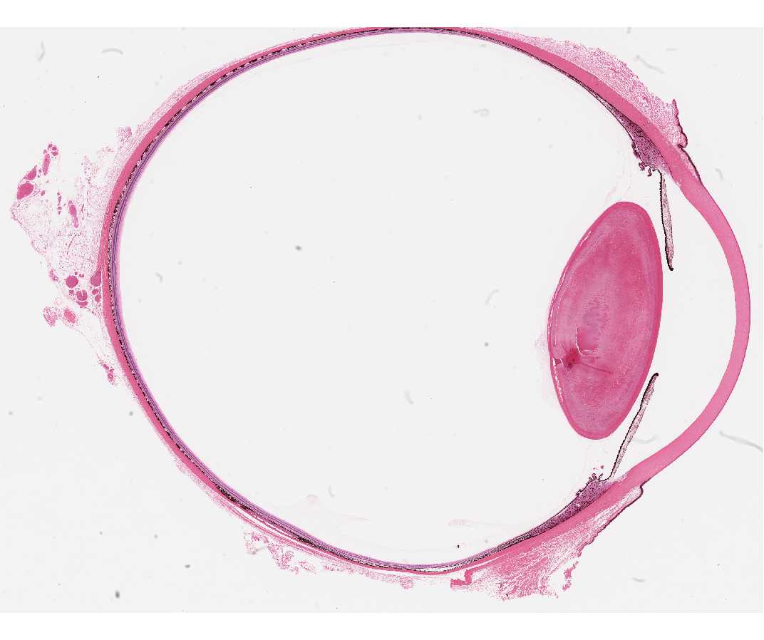
Light microscopic images showing the structure of the cornea stained... |  Download Scientific Diagram
Light microscopic images showing the structure of the cornea stained... | Download Scientific Diagram

Corneal confocal microscopy detects small nerve fibre damage in patients  with painful diabetic neuropathy | Scientific Reports
Corneal confocal microscopy detects small nerve fibre damage in patients with painful diabetic neuropathy | Scientific Reports

Figure 1 from The normal and abnormal human corneal epithelial surface: a  scanning electron microscope study. | Semantic Scholar
Figure 1 from The normal and abnormal human corneal epithelial surface: a scanning electron microscope study. | Semantic Scholar

CS4 Corneal Confocal Microscope - YouTube

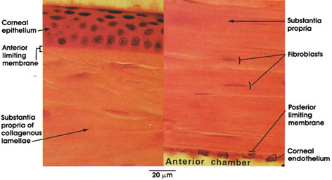
Histology Laboratory Manual

Auricularia cornea, under the microscope | showing just how … | Flickr

Light microscopy of control and KC corneas stained with PAS and Mayer's...  | Download Scientific Diagram
Light microscopy of control and KC corneas stained with PAS and Mayer's... | Download Scientific Diagram

Light microscopic examination of cornea stained with hematoxylin-eosin....  | Download Scientific Diagram
Light microscopic examination of cornea stained with hematoxylin-eosin.... | Download Scientific Diagram

In vivo confocal microscopy of the human cornea | British Journal of  Ophthalmology
In vivo confocal microscopy of the human cornea | British Journal of Ophthalmology

Confocal microscopy in cornea guttata and Fuchs' endothelial dystrophy |  British Journal of Ophthalmology
Confocal microscopy in cornea guttata and Fuchs' endothelial dystrophy | British Journal of Ophthalmology

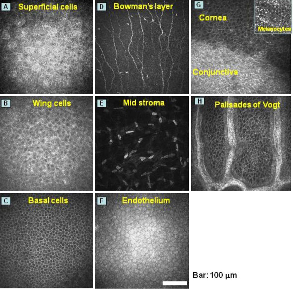
Morphological evaluation of normal human corneal epithelium - Ehlers - 2010  - Acta Ophthalmologica - Wiley Online Library
Morphological evaluation of normal human corneal epithelium - Ehlers - 2010 - Acta Ophthalmologica - Wiley Online Library

Use Specular Microscopy to Diagnose Corneal Disease

Anatomy Atlases: Atlas of Microscopic Anatomy: Section 1 - Cells

Molecular Vision: Chikama, Mol Vis 2009; 15:2896-2904. Figure 1

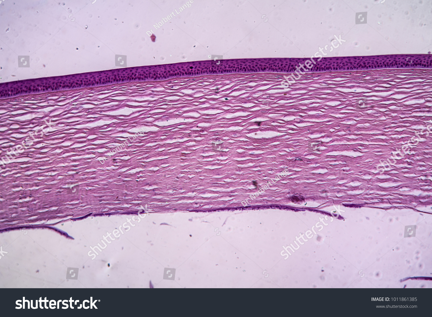
Cornea Eye Tissue Under Microscope 100x Stock Photo 1011861385 |  Shutterstock
Cornea Eye Tissue Under Microscope 100x Stock Photo 1011861385 | Shutterstock

Scanning Electron Microscopy of the Human Cornea

Scanning Electron Microscopy of the Human Cornea

Human Structure Virtual Microscopy

Corneal confocal microscopy identifies small fibre damage and progression  of diabetic neuropathy | Scientific Reports
Corneal confocal microscopy identifies small fibre damage and progression of diabetic neuropathy | Scientific Reports
Cornea eye tissue under the microscope 100x Stock Photo - Alamy

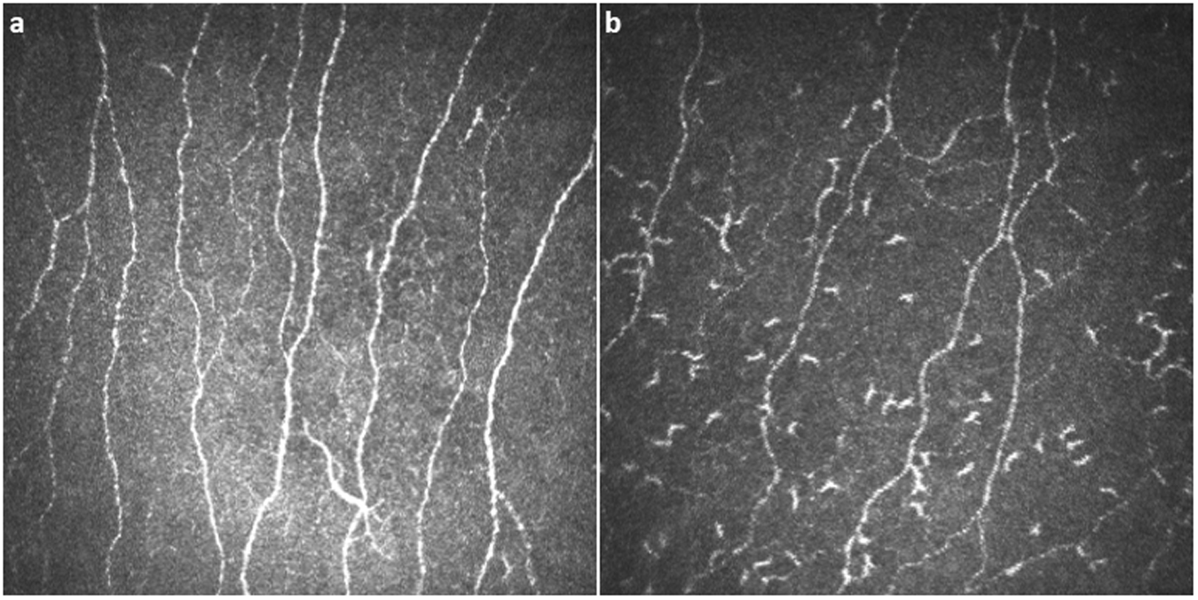
Corneal confocal microscopy detects corneal nerve damage and increased  dendritic cells in Fabry disease | Scientific Reports
Corneal confocal microscopy detects corneal nerve damage and increased dendritic cells in Fabry disease | Scientific Reports

Cornea, Section Microscope Slide - Walmart.com