Home

prin intermediul mecanism rupt human cheak cell microscope cețos Ridica frunze înfrângere

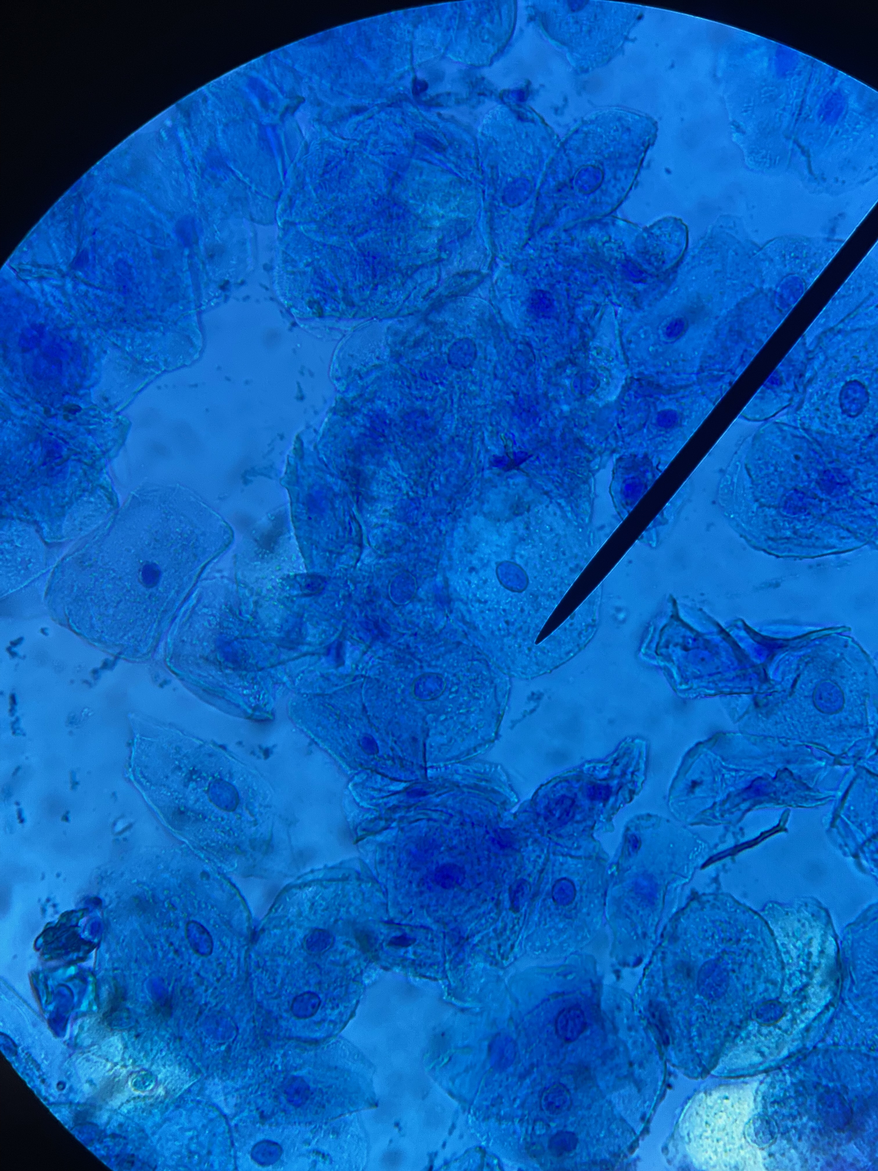
Human Cheek Cell Station

Hands-On Activity: Human Cheek Cells | Science Supply

A human cheek cell and a spongy mesophyll cell are examined under a  microscope. Which structure are seen in both cells? - Quora
A human cheek cell and a spongy mesophyll cell are examined under a microscope. Which structure are seen in both cells? - Quora

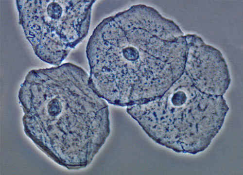
Buccal Epithelial Cells | Nikon's MicroscopyU

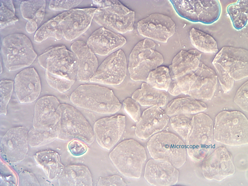
Microscope World Blog: Cheek Cells under Phase Contrast Microscope

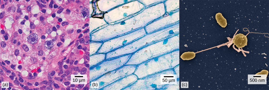
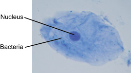
Introduction – Biology

What organelles would be visible in a cheek cell? Why? - Quora

Cell Structure
Cell Structure

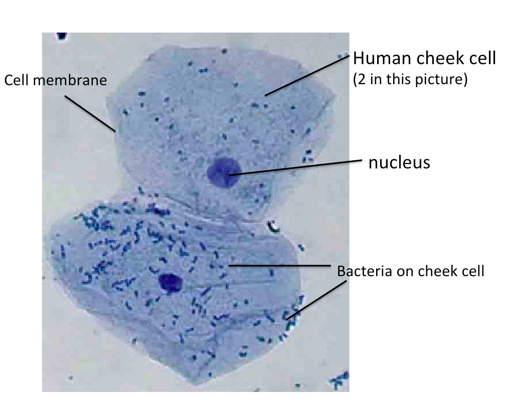
File:Human cheek cell.jpg - Wikimedia Commons

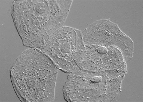
File:Microscopic Image of Human Cheek Cells.jpg - Wikimedia Commons

Draw three types of cells (Cheek cell, Red blood cell, Elodea). Make sure  that you have also labeled these drawings with the cell structures that you  can see (nucleus, cell membrane, cell
Draw three types of cells (Cheek cell, Red blood cell, Elodea). Make sure that you have also labeled these drawings with the cell structures that you can see (nucleus, cell membrane, cell

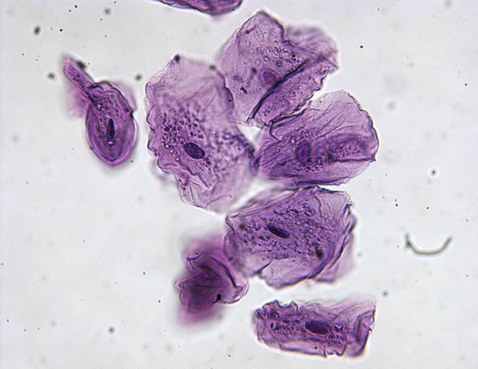
Human Cheek Epithelial Cells | Olympus LS

Cheek cells 400X: General Biology Lab II: Loyola University Chicago

United Scientific Supplies 500-7"Human Cheek Cells" Prepared Microscope  Slide: Amazon.com: Industrial & Scientific
United Scientific Supplies 500-7"Human Cheek Cells" Prepared Microscope Slide: Amazon.com: Industrial & Scientific

Solved Using this table from the Size Estimation module, | Chegg.com

Human Cheek Epithelial Cells. the Tissue that Lines the Inside of the Mouth  is Known As the Basal Mucosa and is Composed of Stock Image - Image of  epithelial, diagnosis: 167106771
Human Cheek Epithelial Cells. the Tissue that Lines the Inside of the Mouth is Known As the Basal Mucosa and is Composed of Stock Image - Image of epithelial, diagnosis: 167106771

Human Cheek - Experiments on Microscopes 4 Schools

Looking at cheek cells under a microscope. | Fun science, Human body,  Things under a microscope
Looking at cheek cells under a microscope. | Fun science, Human body, Things under a microscope

Human cheek cell on PCS. (a) Bright field microscopy. (b) Phase... |  Download Scientific Diagram
Human cheek cell on PCS. (a) Bright field microscopy. (b) Phase... | Download Scientific Diagram

What is the shape of cheek cells and how can you find out the shape of cheek  cells? - Quora
What is the shape of cheek cells and how can you find out the shape of cheek cells? - Quora

Human Cheek Epidermis

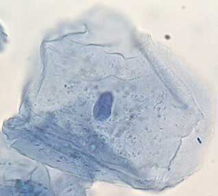
The Human Cheek Cell

Cheek Epithelial Cell Practical Experiment - YouTube

Difference Between Onion Cell and Human Cheek Cell - Pediaa.Com