Home

aparţine Piscina Convoca optovue mati Optimist fotografie Milestone

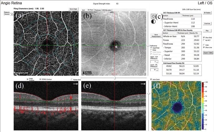
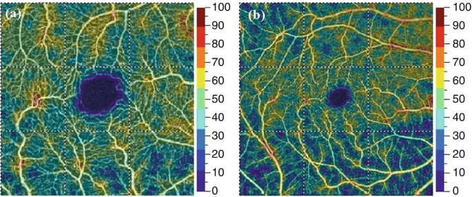
VASCULAR COMPLEXITY ANALYSIS IN OPTICAL COHERENCE TOMOGRAPHY ANGIOGRAPHY OF  DIABETIC RETINOPATHY
VASCULAR COMPLEXITY ANALYSIS IN OPTICAL COHERENCE TOMOGRAPHY ANGIOGRAPHY OF DIABETIC RETINOPATHY

OCT in Glaucoma | SpringerLink

Decision Making in Glaucoma: When to pull the trigger” COPE #41665-GL  Disclosures Risk Factors for Glaucoma Glaucoma Primar
Decision Making in Glaucoma: When to pull the trigger” COPE #41665-GL Disclosures Risk Factors for Glaucoma Glaucoma Primar
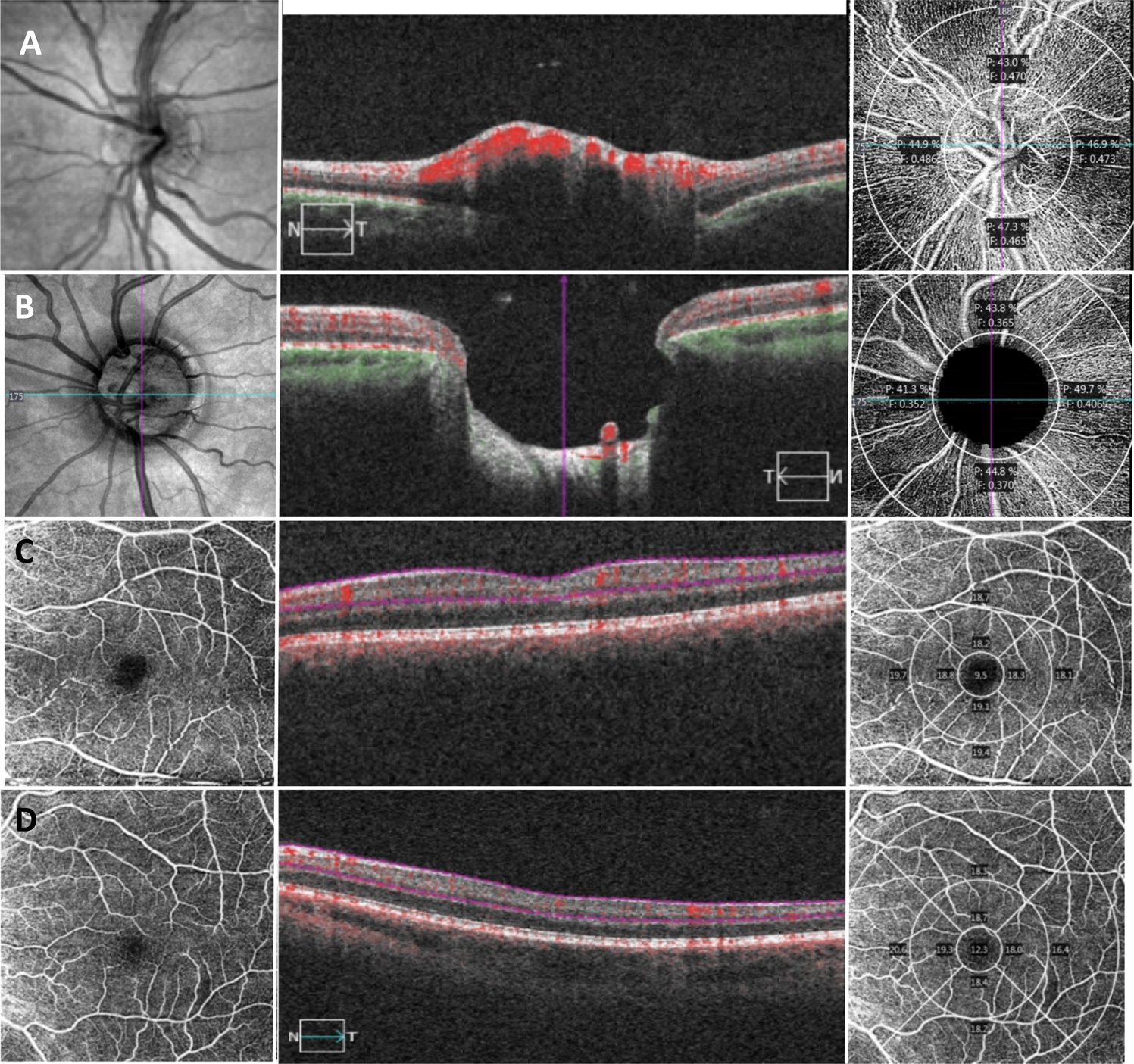
Quantitative features for objective assessment of OCT angiography - Book  chapter - IOPscience
Quantitative features for objective assessment of OCT angiography - Book chapter - IOPscience

OCT in Glaucoma | SpringerLink

Peripapillary and macular vascular parameters by optical coherence  tomography angiography in primary congenital glaucoma | Eye
Peripapillary and macular vascular parameters by optical coherence tomography angiography in primary congenital glaucoma | Eye

Anterior segment optical coherence tomography - ScienceDirect

Validation of automated artificial intelligence segmentation of optical  coherence tomography images | PLOS ONE
Validation of automated artificial intelligence segmentation of optical coherence tomography images | PLOS ONE
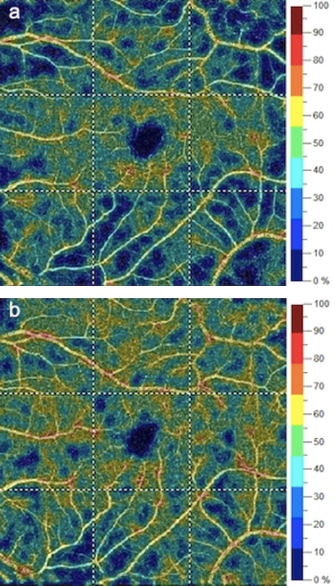
OCT in Glaucoma | SpringerLink

Segmentation of anterior segment boundaries in swept source OCT images -  ScienceDirect
Segmentation of anterior segment boundaries in swept source OCT images - ScienceDirect

Anterior segment optical coherence tomography - ScienceDirect

Changes in optic nerve head and macula optical coherence tomography  angiography parameters before and after trabeculectomy | SpringerLink
Changes in optic nerve head and macula optical coherence tomography angiography parameters before and after trabeculectomy | SpringerLink
Strengthen
Strengthen

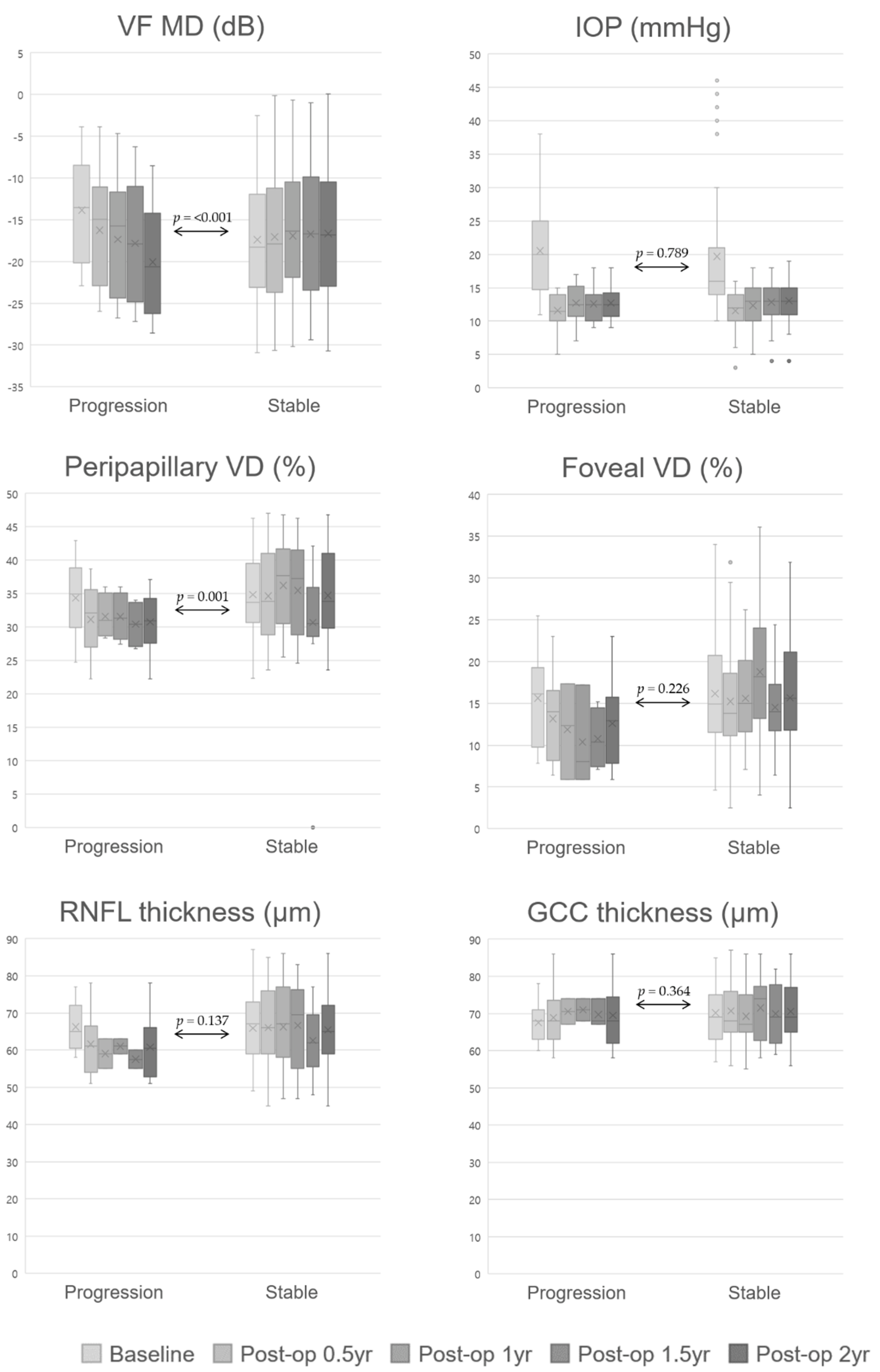
JCM | Free Full-Text | Changes in Peripapillary and Macular Vessel  Densities and Their Relationship with Visual Field Progression after  Trabeculectomy | HTML
JCM | Free Full-Text | Changes in Peripapillary and Macular Vessel Densities and Their Relationship with Visual Field Progression after Trabeculectomy | HTML

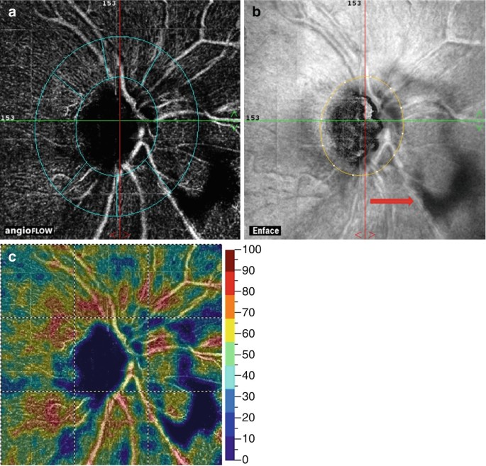
Optic Disc Microvasculature Dropout in Glaucoma Detected by Swept-Source  Optical Coherence Tomography Angiography - American Journal of Ophthalmology
Optic Disc Microvasculature Dropout in Glaucoma Detected by Swept-Source Optical Coherence Tomography Angiography - American Journal of Ophthalmology

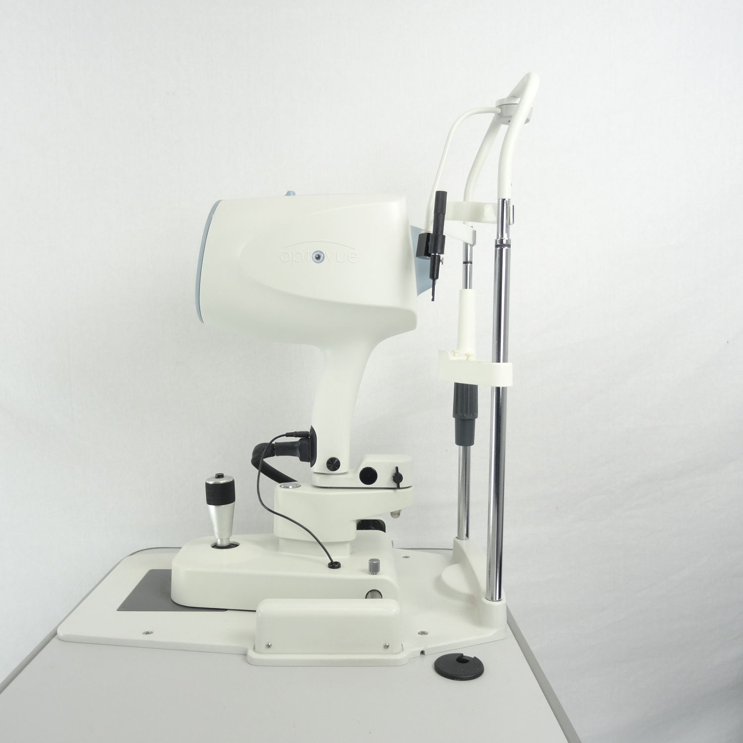
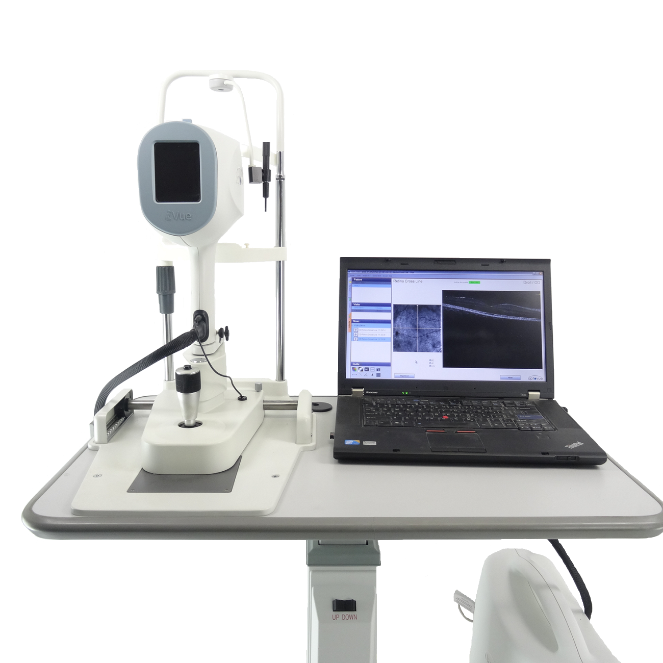
OPTOVUE OCT i VUE - Medlikim

Relationship between the rate of change in lamina cribrosa depth and the  rate of retinal nerve fiber layer thinning following glaucoma surgery |  PLOS ONE
Relationship between the rate of change in lamina cribrosa depth and the rate of retinal nerve fiber layer thinning following glaucoma surgery | PLOS ONE

CE Certificate Blank 2018

OPTOVUE OCT i VUE - Medlikim

INNOVATION IN RETINAL IMAGING: OPPORTUNITIES & CHALLENGES - History o…

PDF) Association between Optical Coherence Tomography Measurements and  Clinical Parameters in Idiopathic Intracranial Hypertension
PDF) Association between Optical Coherence Tomography Measurements and Clinical Parameters in Idiopathic Intracranial Hypertension