Home

rănit Botanică Făcut sămi amintesc plant cuticle glandular microscope Expres Danemarca

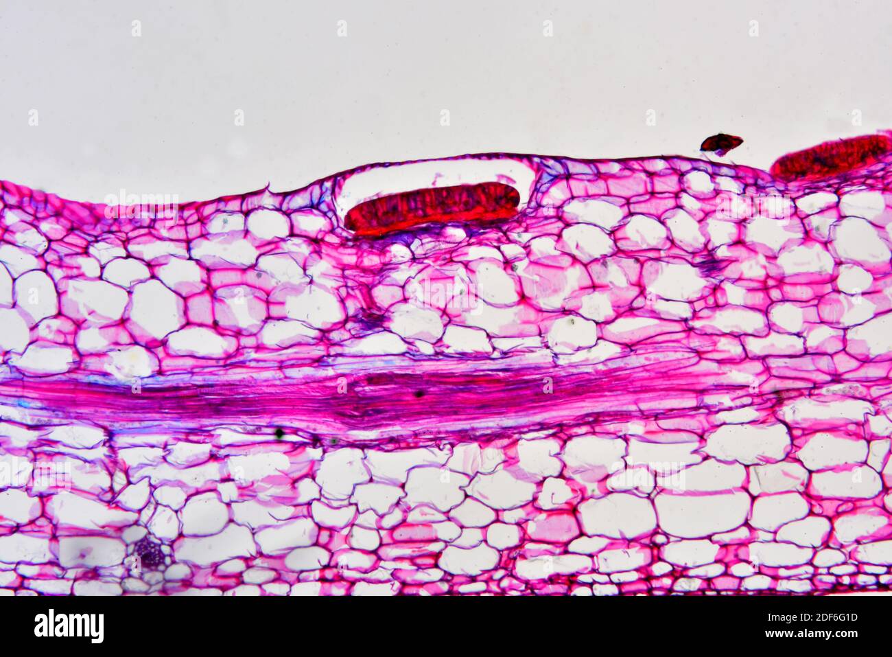
File:Cuticle of leaf under microscope.JPG - Wikimedia Commons

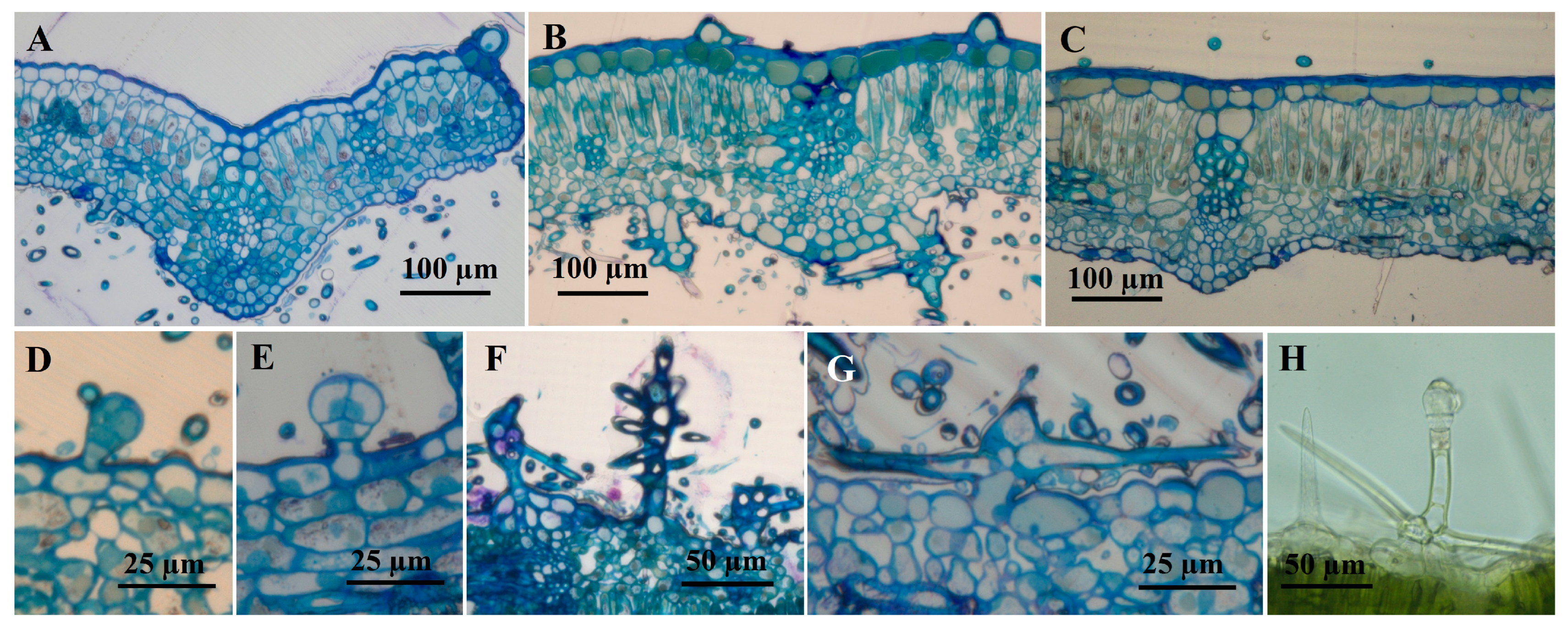
Structures and distributions of glandular trichomes in Vitis. (A–C)... |  Download Scientific Diagram
Structures and distributions of glandular trichomes in Vitis. (A–C)... | Download Scientific Diagram

SciELO - Brasil - Morphology and histochemistry of glandular trichomes in  <i>Hyptis villosa</i> Pohl ex Benth. (Lamiaceae) and differential labeling  of cytoskeletal elements Morphology and histochemistry of glandular  trichomes in <i>Hyptis villosa</i>
SciELO - Brasil - Morphology and histochemistry of glandular trichomes in <i>Hyptis villosa</i> Pohl ex Benth. (Lamiaceae) and differential labeling of cytoskeletal elements Morphology and histochemistry of glandular trichomes in <i>Hyptis villosa</i>

Leaf epidermal surfaces under optical microscope. 2A: Abaxial surface... |  Download Scientific Diagram
Leaf epidermal surfaces under optical microscope. 2A: Abaxial surface... | Download Scientific Diagram
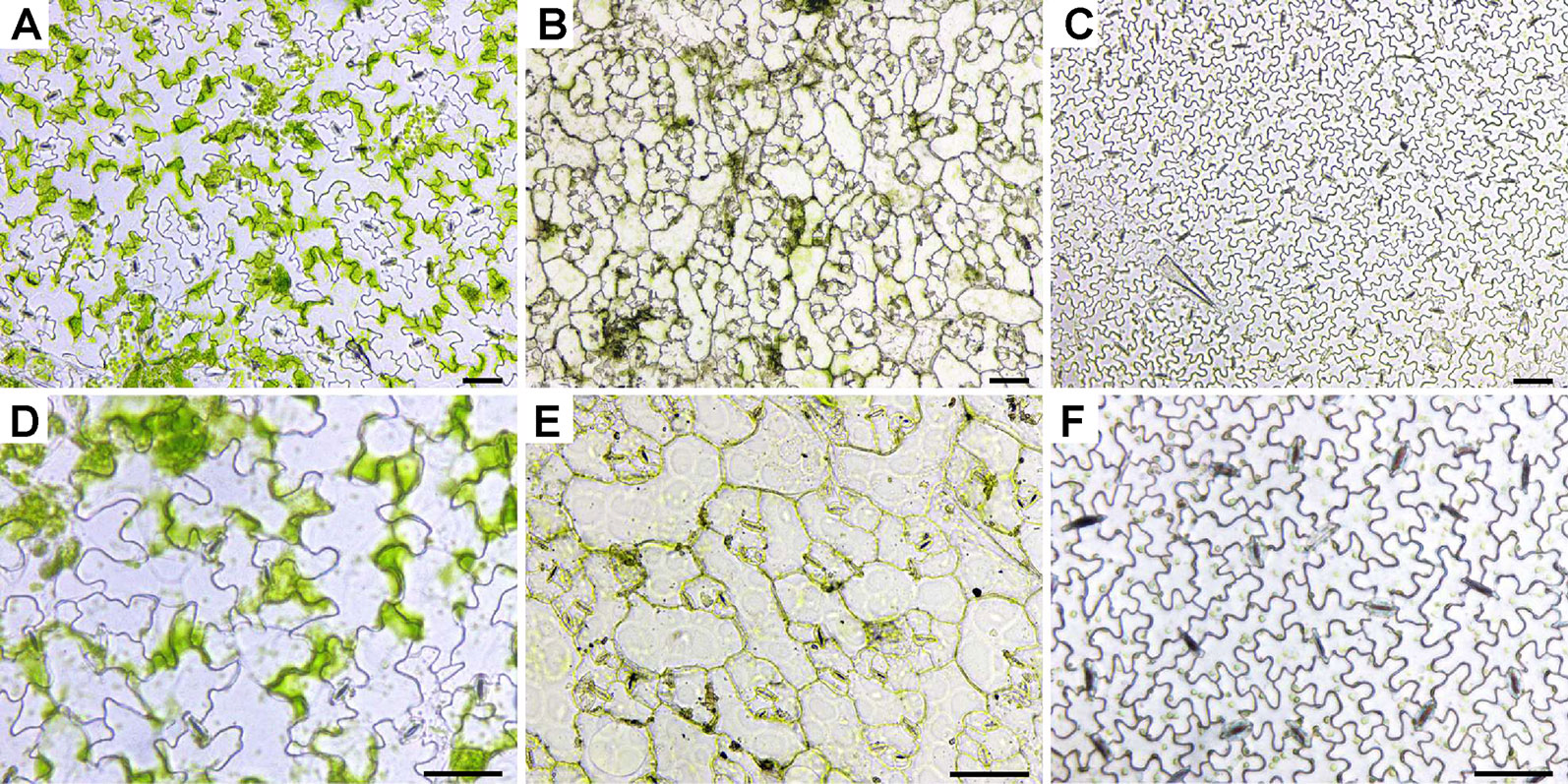
Frontiers | Comparison of Sample Preparation Techniques for Inspection of  Leaf Epidermises Using Light Microscopy and Scanning Electronic Microscopy
Frontiers | Comparison of Sample Preparation Techniques for Inspection of Leaf Epidermises Using Light Microscopy and Scanning Electronic Microscopy

Plant tissues. Protection. Atlas of plant and animal histology.

Comparative anatomy of the vegetative organs of the phanerogams and ferns.  Plant anatomy; Phanerogams; Ferns. 90 CELLULAR TISSUE. and treated with  dissolving reagentsâe. g. alcohol or etherâis intercalated between  cell-membrane and
Comparative anatomy of the vegetative organs of the phanerogams and ferns. Plant anatomy; Phanerogams; Ferns. 90 CELLULAR TISSUE. and treated with dissolving reagentsâe. g. alcohol or etherâis intercalated between cell-membrane and
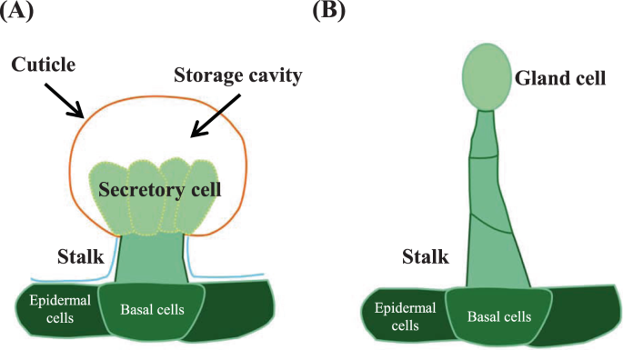
Formation mechanism of glandular trichomes involved in the synthesis and  storage of terpenoids in lavender | BMC Plant Biology | Full Text
Formation mechanism of glandular trichomes involved in the synthesis and storage of terpenoids in lavender | BMC Plant Biology | Full Text
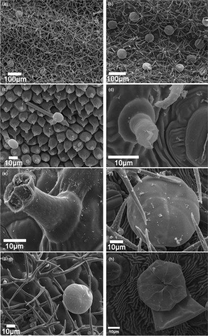
Leaf surface of Rosmarinus officinalis imaged by scanning electron... |  Download Scientific Diagram
Leaf surface of Rosmarinus officinalis imaged by scanning electron... | Download Scientific Diagram

Plant cell microscope hi-res stock photography and images - Page 2 - Alamy

Plants | Free Full-Text | Glandular and Non-Glandular Trichomes from  Phlomis herba-venti subsp. pungens Leaves: Light, Confocal, and Scanning  Electron Microscopy and Histochemistry of the Secretory Products
Plants | Free Full-Text | Glandular and Non-Glandular Trichomes from Phlomis herba-venti subsp. pungens Leaves: Light, Confocal, and Scanning Electron Microscopy and Histochemistry of the Secretory Products

Plant tissues. Protection. Trichomes. Atlas of plant and animal tissues.

Genetic Control of Glandular Trichome Development: Trends in Plant Science

Secretory structures in the leaves of Hibiscus sabdariffa L. - ScienceDirect

Morphology of the capitate glandular trichomes from the staminode of... |  Download Scientific Diagram
Morphology of the capitate glandular trichomes from the staminode of... | Download Scientific Diagram

Plants | Free Full-Text | Glandular and Non-Glandular Trichomes from  Phlomis herba-venti subsp. pungens Leaves: Light, Confocal, and Scanning  Electron Microscopy and Histochemistry of the Secretory Products
Plants | Free Full-Text | Glandular and Non-Glandular Trichomes from Phlomis herba-venti subsp. pungens Leaves: Light, Confocal, and Scanning Electron Microscopy and Histochemistry of the Secretory Products

Lucilia nitens Less., Asteraceae, leaf. A: scanning electron microscopy...  | Download Scientific Diagram
Lucilia nitens Less., Asteraceae, leaf. A: scanning electron microscopy... | Download Scientific Diagram

Morphological investigation of glandular hairs on Drosera capensis leaves  with an ultrastructural study of the sessile glands
Morphological investigation of glandular hairs on Drosera capensis leaves with an ultrastructural study of the sessile glands
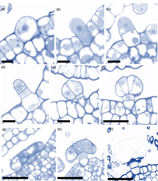
Formation mechanism of glandular trichomes involved in the synthesis and  storage of terpenoids in lavender | BMC Plant Biology | Full Text
Formation mechanism of glandular trichomes involved in the synthesis and storage of terpenoids in lavender | BMC Plant Biology | Full Text

Convergence of glandular trichome morphology and chemistry in two montane  monkeyflower species | bioRxiv
Convergence of glandular trichome morphology and chemistry in two montane monkeyflower species | bioRxiv
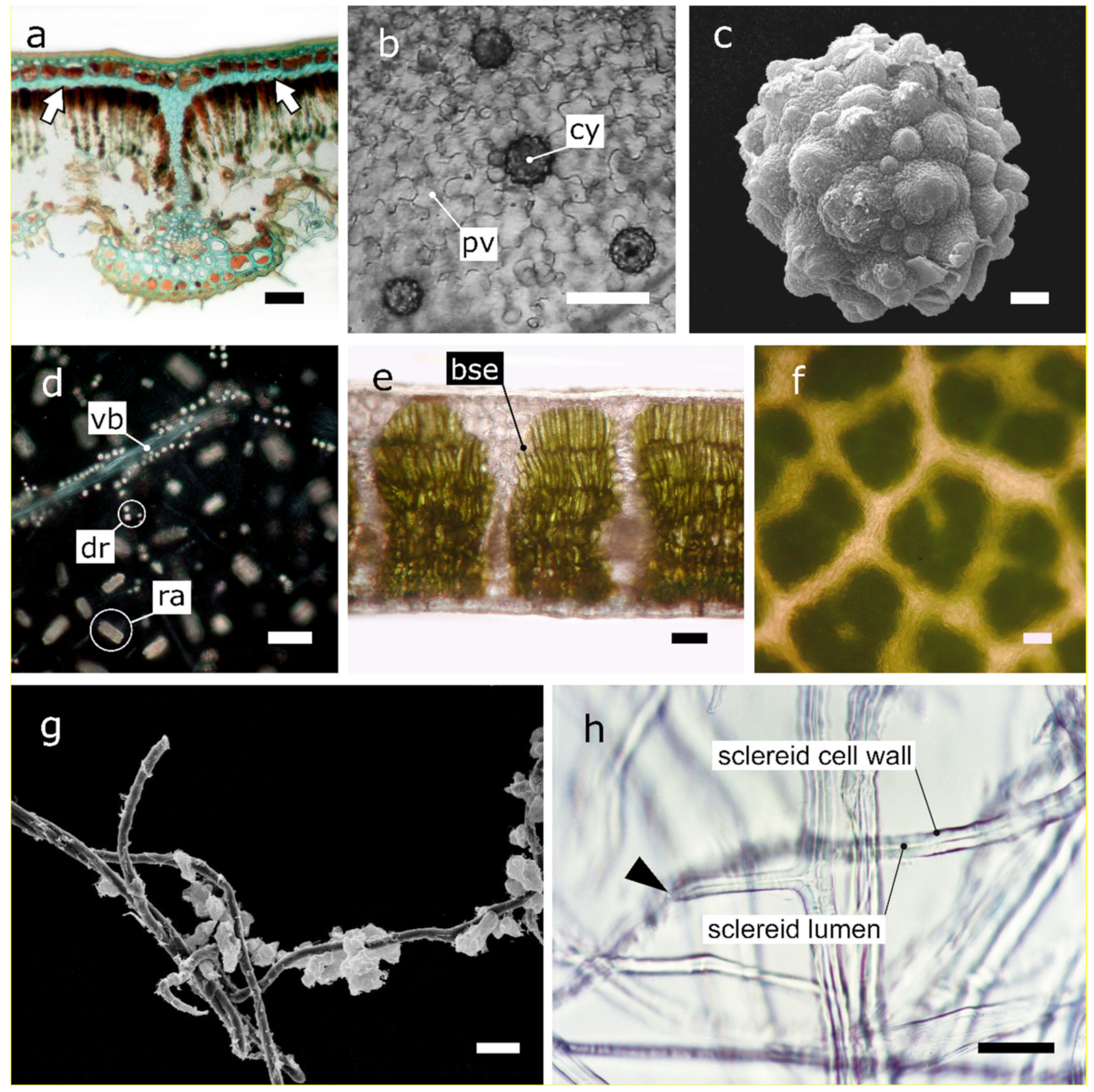
Plants | Free Full-Text | The Optical Properties of Leaf Structural  Elements and Their Contribution to Photosynthetic Performance and  Photoprotection
Plants | Free Full-Text | The Optical Properties of Leaf Structural Elements and Their Contribution to Photosynthetic Performance and Photoprotection

Light microscopy observations of a capitate gland with a single head... |  Download Scientific Diagram
Light microscopy observations of a capitate gland with a single head... | Download Scientific Diagram
Glandular trichomes: new focus on horticultural crops | Horticulture  Research
Glandular trichomes: new focus on horticultural crops | Horticulture Research

Genetic Control of Glandular Trichome Development: Trends in Plant Science

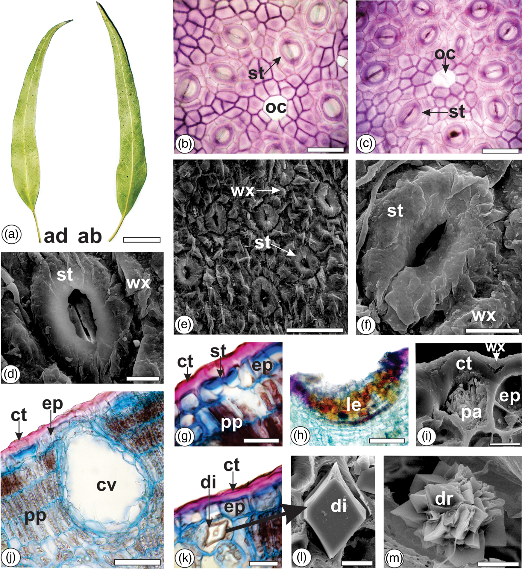
Light and Scanning Electron Microscopy, Energy Dispersive X-Ray  Spectroscopy, and Histochemistry of Eucalyptus tereticornis | Microscopy  and Microanalysis | Cambridge Core
Light and Scanning Electron Microscopy, Energy Dispersive X-Ray Spectroscopy, and Histochemistry of Eucalyptus tereticornis | Microscopy and Microanalysis | Cambridge Core