Home

A juca jocuri sportive Nuc persecuţie skull mri lamina anatomy Uimire busolă te enervează

Brain: Atlas of human anatomy with MRI - e-Anatomy

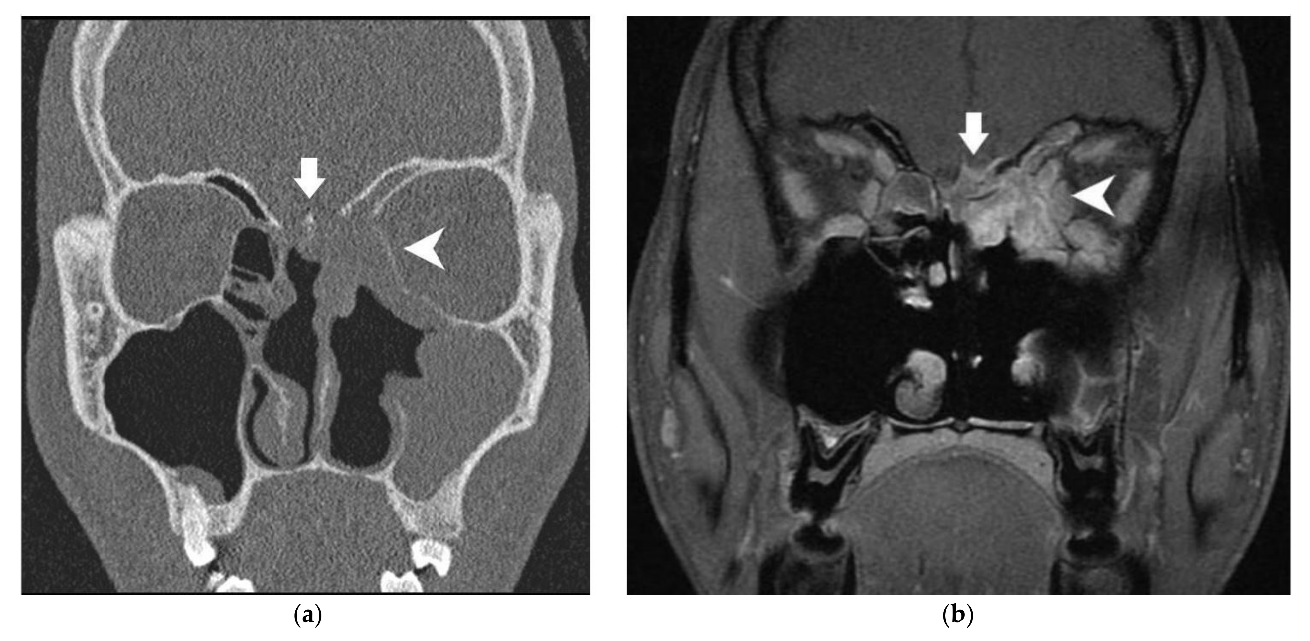
Cancers | Free Full-Text | Imaging of Skull Base and Orbital Invasion in  Sinonasal Cancer: Correlation with Histopathology | HTML
Cancers | Free Full-Text | Imaging of Skull Base and Orbital Invasion in Sinonasal Cancer: Correlation with Histopathology | HTML
MIDA: A Multimodal Imaging-Based Detailed Anatomical Model of the Human Head  and Neck | PLOS ONE
MIDA: A Multimodal Imaging-Based Detailed Anatomical Model of the Human Head and Neck | PLOS ONE

Cross-Sectional Computed Tomography and Magnetic Resonance Imaging Atlas of  the Skull Base | Radiology Key
Cross-Sectional Computed Tomography and Magnetic Resonance Imaging Atlas of the Skull Base | Radiology Key

Lesions of the Hypothalamus: MR Imaging Diagnostic Features | RadioGraphics

Twenty-Five Diagnoses on Midline Images of the Brain: From Fetus to Child  to Adult | RadioGraphics
Twenty-Five Diagnoses on Midline Images of the Brain: From Fetus to Child to Adult | RadioGraphics

Skull Base–related Lesions at Routine Head CT from the Emergency  Department: Pearls, Pitfalls, and Lessons Learned | RadioGraphics
Skull Base–related Lesions at Routine Head CT from the Emergency Department: Pearls, Pitfalls, and Lessons Learned | RadioGraphics

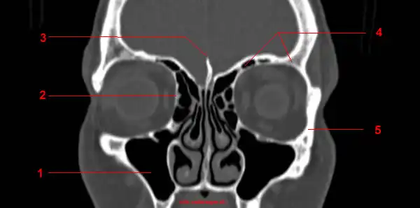
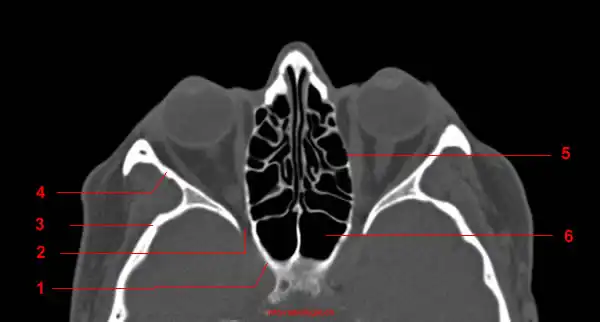
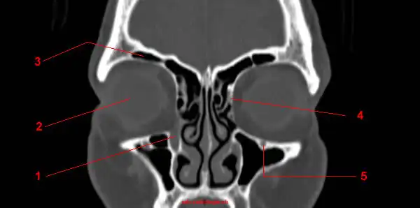
CT of the Orbit: anatomy - W-Radiology

Normal anatomy of the base of the skull, orbit, pituitary and cranial  nerves | Practical Neurology
Normal anatomy of the base of the skull, orbit, pituitary and cranial nerves | Practical Neurology

Imaging review of the anterior skull base - Olivia Francies, Levan  Makalanda, Dimitris Paraskevopolous, Ashok Adams, 2018
Imaging review of the anterior skull base - Olivia Francies, Levan Makalanda, Dimitris Paraskevopolous, Ashok Adams, 2018

CT of the Orbit: anatomy - W-Radiology

A) Sagittal, (B) coronal, and (C) axial T1 weighted magnetic resonance... |  Download Scientific Diagram
A) Sagittal, (B) coronal, and (C) axial T1 weighted magnetic resonance... | Download Scientific Diagram

CT of the Orbit: anatomy - W-Radiology

Skull | Radiology Reference Article | Radiopaedia.org

Brain: Atlas of human anatomy with MRI - e-Anatomy

Hypothalamus - W-Radiology

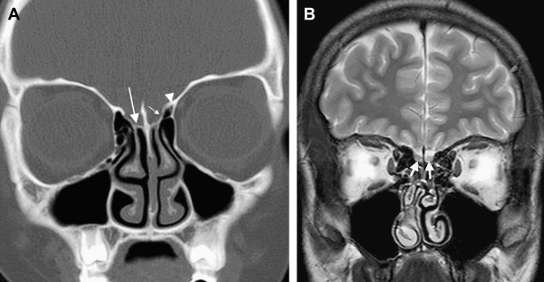
Skull base tumours part I: Imaging technique, anatomy and anterior skull  base tumours - European Journal of Radiology
Skull base tumours part I: Imaging technique, anatomy and anterior skull base tumours - European Journal of Radiology
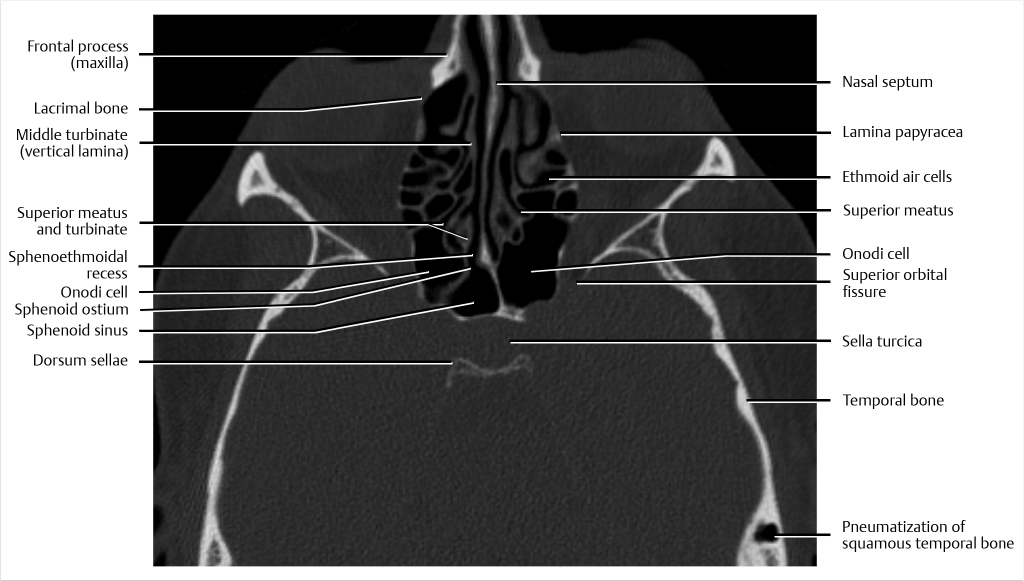
Imaging of the Anterior Skull Base | Radiology Key

In-vivo magnetic resonance imaging (MRI) of laminae in the human cortex -  ScienceDirect
In-vivo magnetic resonance imaging (MRI) of laminae in the human cortex - ScienceDirect

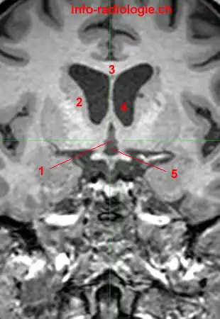
Lamina terminalis | Radiology Reference Article | Radiopaedia.org

Sagittal midline of the brain - normal anatomy | Radiology Case |  Radiopaedia.org
Sagittal midline of the brain - normal anatomy | Radiology Case | Radiopaedia.org

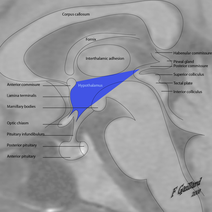
Radiopaedia - Drawing Boundaries of the hypothalamus with translucent MRI -  English labels | AnatomyTOOL
Radiopaedia - Drawing Boundaries of the hypothalamus with translucent MRI - English labels | AnatomyTOOL